Human body dissection in 3D at WSU
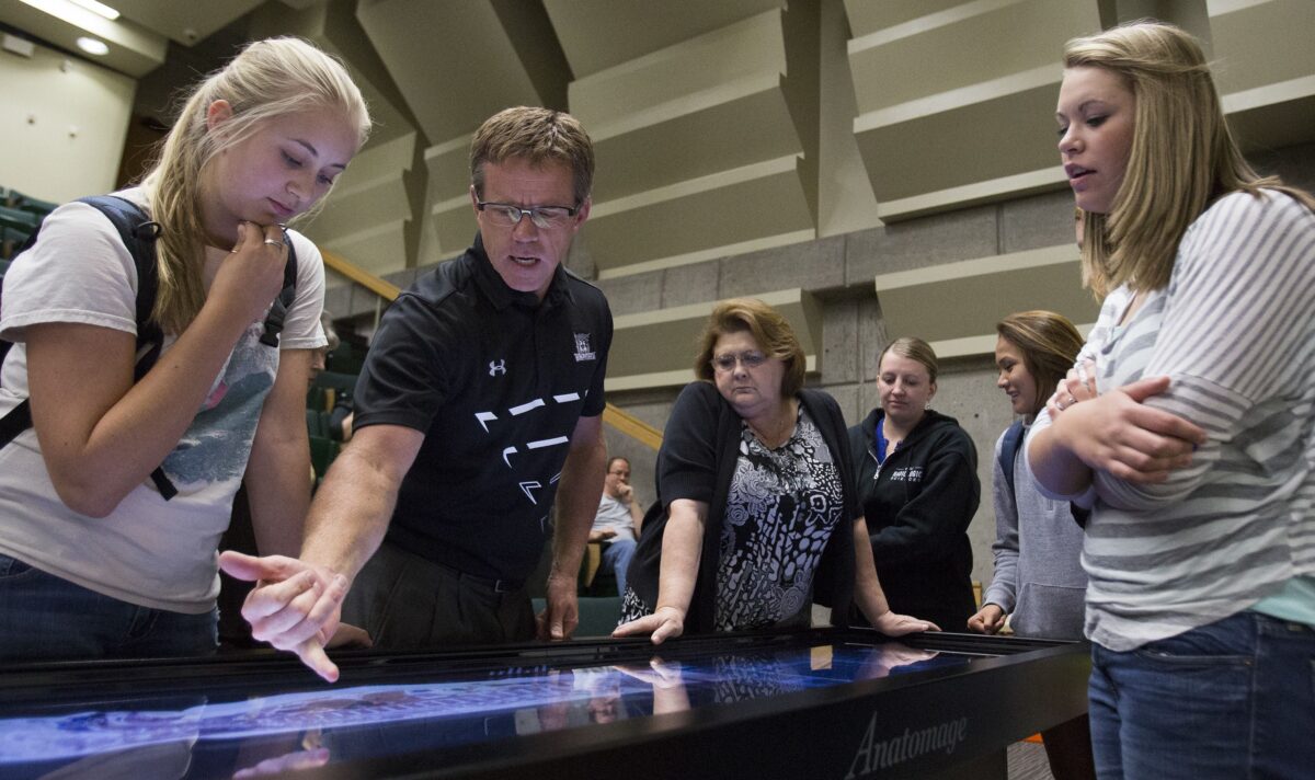
Chair of the Department of Heath Sciences Kraig Chugg demonstrated to faculty and students the capabilities of Weber State University's new Anatomage table at the Marriott Allied Health Building at Weber State University on Thursday, September 11, 2014.
Chair of the Department of Heath Sciences Kraig Chugg demonstrated to faculty and students the capabilities of Weber State University's new Anatomage table at the Marriott Allied Health Building at Weber State University on Thursday, September 11, 2014. He compared the table to a plasticized cadaver, which will both be used to teach students.
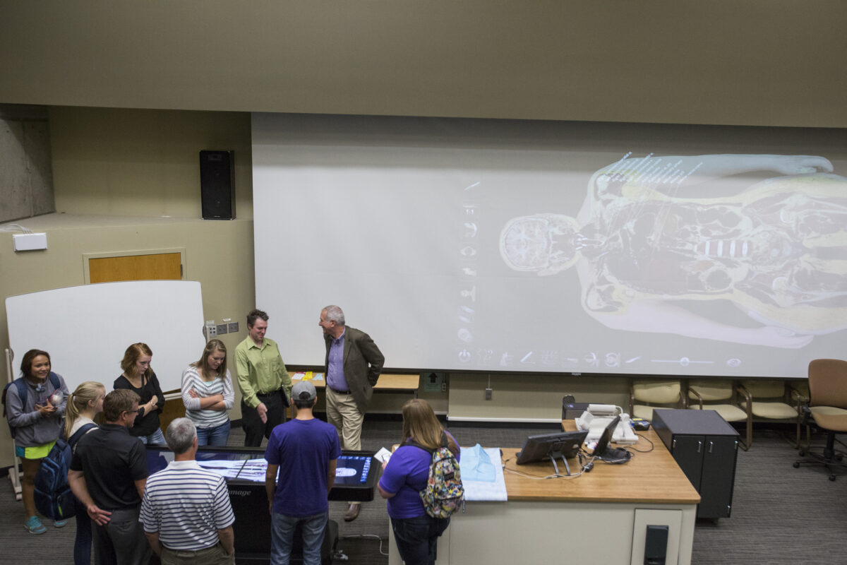
Professor Jim Hutchins, Ph.D, center, talks with faculty and students as they are able to try Weber State University's new Anatomage table at the Marriott Allied Health Building at Weber State University on Thursday, September 11, 2014.
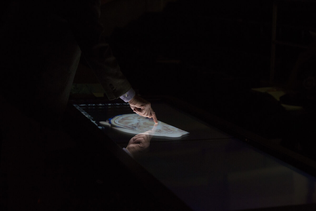
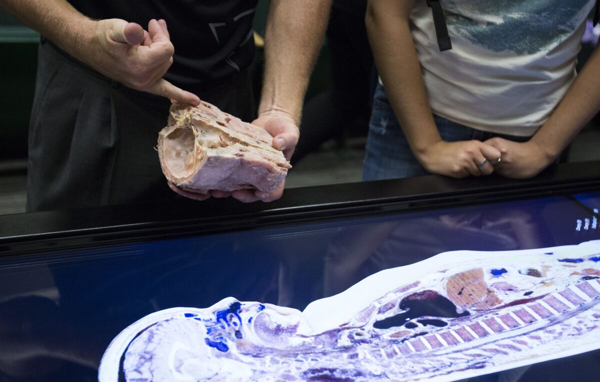
Professor Jim Hutchins, Ph.D, points to areas of the brain on Weber State University's new Anatomage table at the Marriott Allied Health Building at Weber State University on Thursday, September 11, 2014. Similar to smart devices the images can be zoomed in, zoomed out, turned, and moved just with one's finger tips.
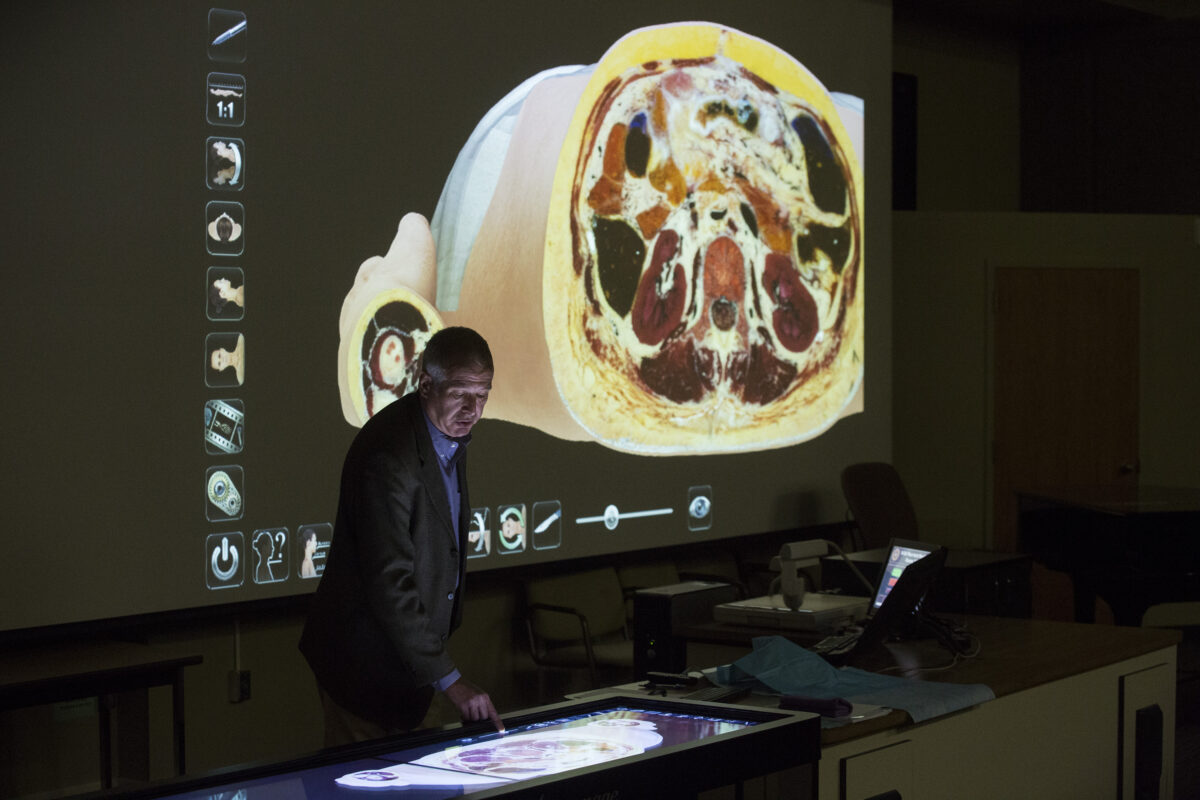
Professor Jim Hutchins, Ph.D, demonstrates to faculty and students Weber State University's new Anatomage table at the Marriott Allied Health Building at Weber State University on Thursday, September 11, 2014. By moving a slider you can view deeper or shallower through the layers the human body.
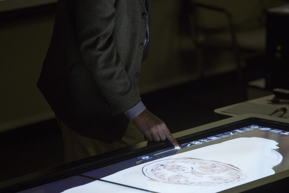
Professor Jim Hutchins, Ph.D, demonstrates to faculty and students Weber State University's new Anatomage table at the Marriott Allied Health Building at Weber State University on Thursday, September 11, 2014. By moving a slider you can view deeper or shallower through the layers the human body.
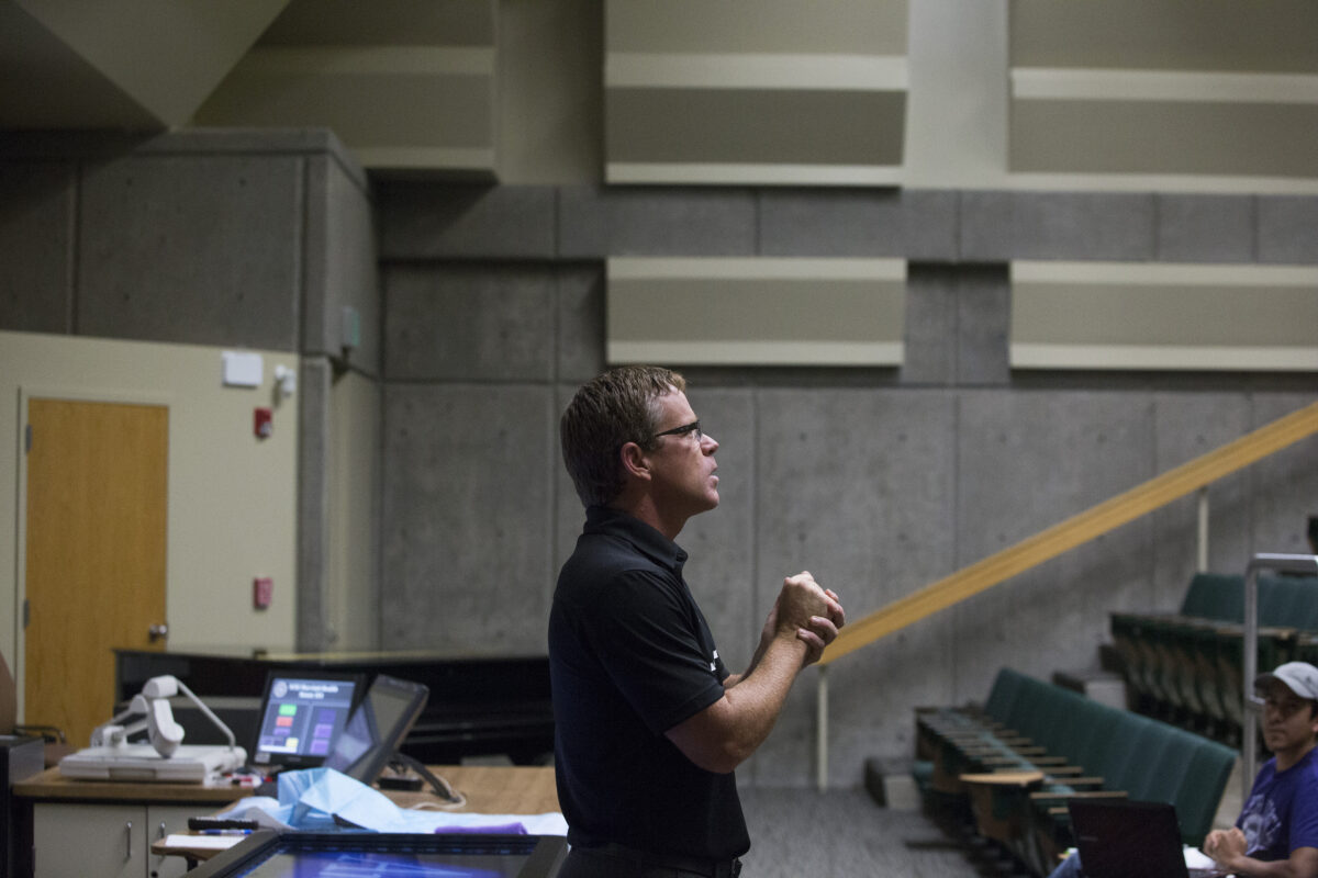
Chair of the Department of Heath Sciences Kraig Chugg spoke during a demonstration to faculty and students of Weber State University's new Anatomage table at the Marriott Allied Health Building at Weber State University on Thursday, September 11, 2014.
OGDEN — With the swipe of two fingers, Weber State University student Nurivan Dominguez delved deep inside a human body, dissecting it from head to toe.
Needless to say, he was a little more than impressed by what he saw.
“I am completely amazed,” he said. “I’ve never seen anything like this before. It’s just incredible.”
A virtual human cadaver patient lay embedded inside a 6-foot table, allowing students to investigate its anatomy in 3D images, rotating and slicing both soft and hard tissue. Then, with a single slide of the finger, the entire body was instantly restored.
The table, called Anatomage, is the latest interactive teaching tool at the University’s Health Science Lab. The high-tech device is complete with virtual lifesize patients who have donated their bodies to science, touch screen navigation and computer software that allows for a deep look into every organ, bone and blood vessel from head to toe.
The touch screen has two LCD screens side by side which allow its users to view the body both inside the table and on a large screen in the classroom. The cadavers can be virtually rotated and dissected in numerous ways, allowing the viewer to see everything, from the inside of the brain all the way down to the tips of the toes. If the student wants to look at an organ unobstructed, they can use an application to dissolve bone, muscle and blood vessels to get a better view.
“This gives a better view than anything in a book or anything in your mind,” Dominguez said. “To me it’s the perfect tool for students, especially those who are looking forward to a career in surgery or another type of medicine.”
The cadaver donors, one male and one female, come from the Visible Human Project which is run by the U.S. National Library of Medicine, said department chairman Kraig Chugg.
The Visible Human Project got its start when 38-year-old Texas murderer Joseph Paul Jernigan, who was executed by lethal injection, agreed to donate his body for scientific research or medical use. His body was encased and frozen in a gelatin and water mixture and then cut in one millimeter intervals, Chugg said. Each slice was photographed and rescanned in high resolution. The female cadaver, who remains anonymous, was cut into slices at .33 millimeter intervals.
WSU uses cadavers from the Korean Visible Project. One of the female cadaver’s showed she suffered complications of an ectopic pregnancy, where the embryo is implanted outside of the uterus. Students were able to digitally remove several organs inside the body, giving way to a much clearer view of the abnormality inside the young woman.
“As you start to remove the skin, muscles, veins and organs, you can see something abnormal on one side of the abdomen,” said Jim Hutchins, a professor in the health and science department. “There’s a bump there that shouldn’t be there.”
As Hutchins continued to zoom in closer, a tiny skeleton was revealed inside on the right side of the woman’s abdomen, showing an ectopic pregnancy, which can be a life threatening condition.
The table, which cost approximately $60,000, was paid for through private donors.